Home

Profesor de escuela Bloquear Migración lps stimulation protocol Ver a través de Arsenal consumo

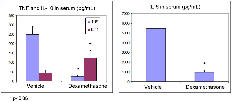
Hooke - Contract Research - Ex vivo whole-blood LPS stimulation

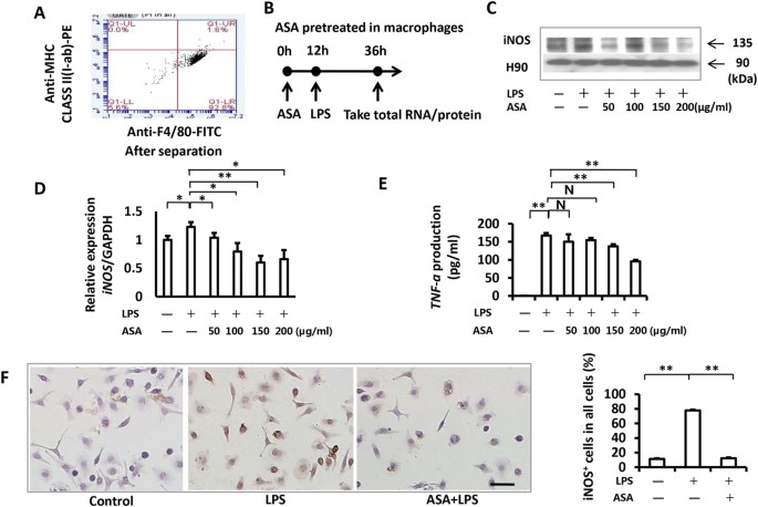
Aspirin inhibits LPS-induced macrophage activation via the NF-κB pathway |  Scientific Reports
Aspirin inhibits LPS-induced macrophage activation via the NF-κB pathway | Scientific Reports

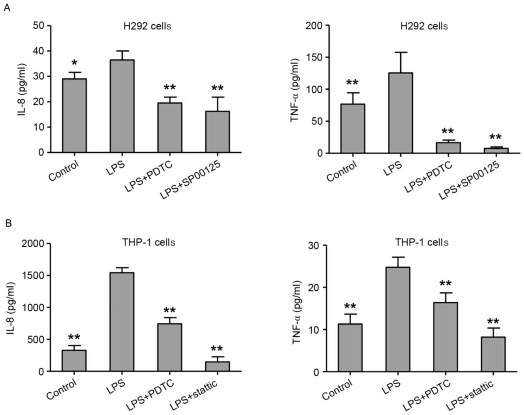
LPS‑induced proinflammatory cytokine expression in human airway epithelial  cells and macrophages via NF‑κB, STAT3 or AP‑1 activation
LPS‑induced proinflammatory cytokine expression in human airway epithelial cells and macrophages via NF‑κB, STAT3 or AP‑1 activation

Steps of ovarian stimulation with DuoStim protocol. Gn = Gonadotrophin,...  | Download Scientific Diagram
Steps of ovarian stimulation with DuoStim protocol. Gn = Gonadotrophin,... | Download Scientific Diagram
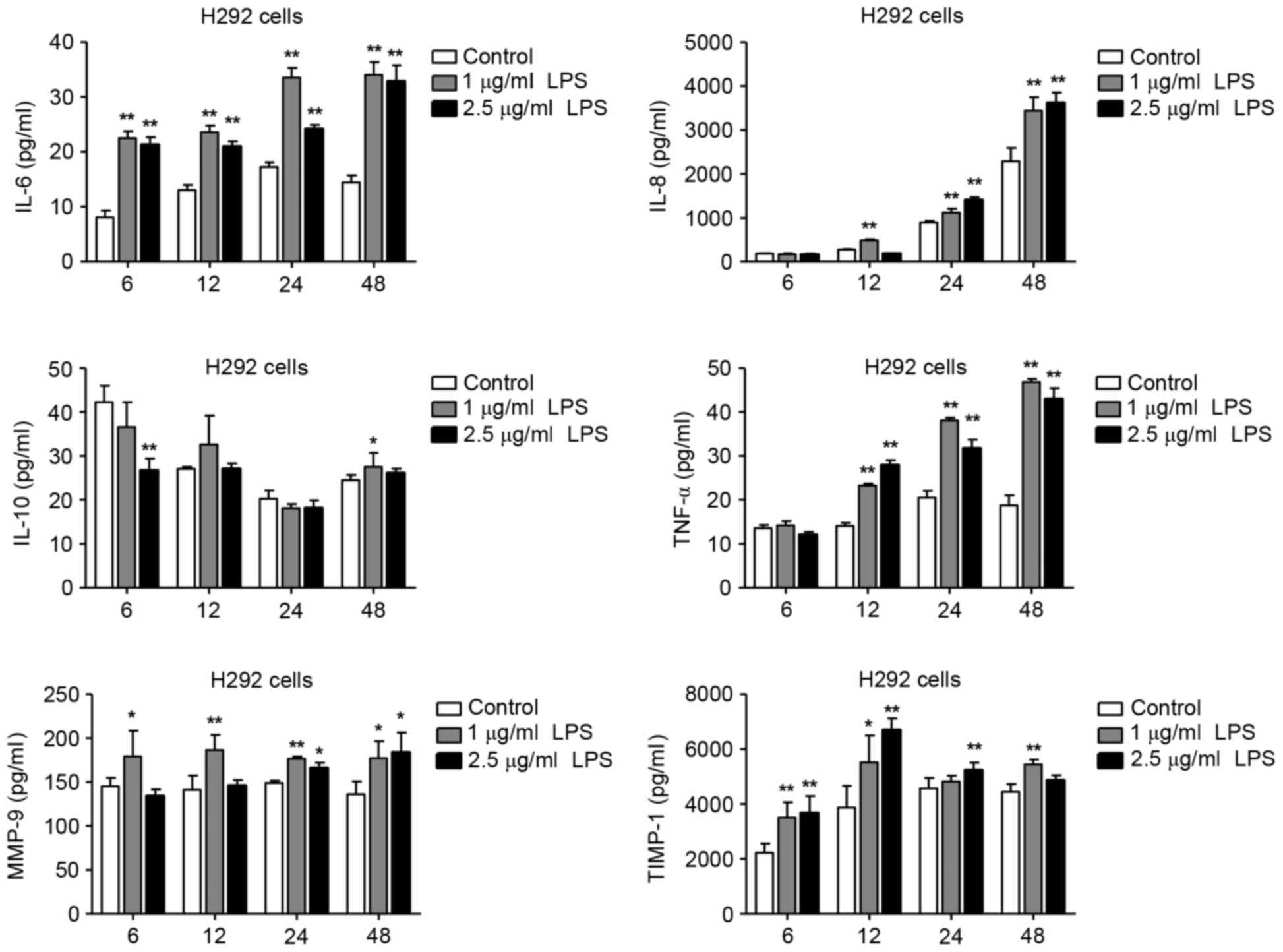
LPS‑induced proinflammatory cytokine expression in human airway epithelial  cells and macrophages via NF‑κB, STAT3 or AP‑1 activation
LPS‑induced proinflammatory cytokine expression in human airway epithelial cells and macrophages via NF‑κB, STAT3 or AP‑1 activation

Repolarization of M(IFNγ+LPS) to M(LPS+IC). (A,B) The protocol used... |  Download Scientific Diagram
Repolarization of M(IFNγ+LPS) to M(LPS+IC). (A,B) The protocol used... | Download Scientific Diagram
Hooke - Contract Research - LPS-Induced Cytokine Production

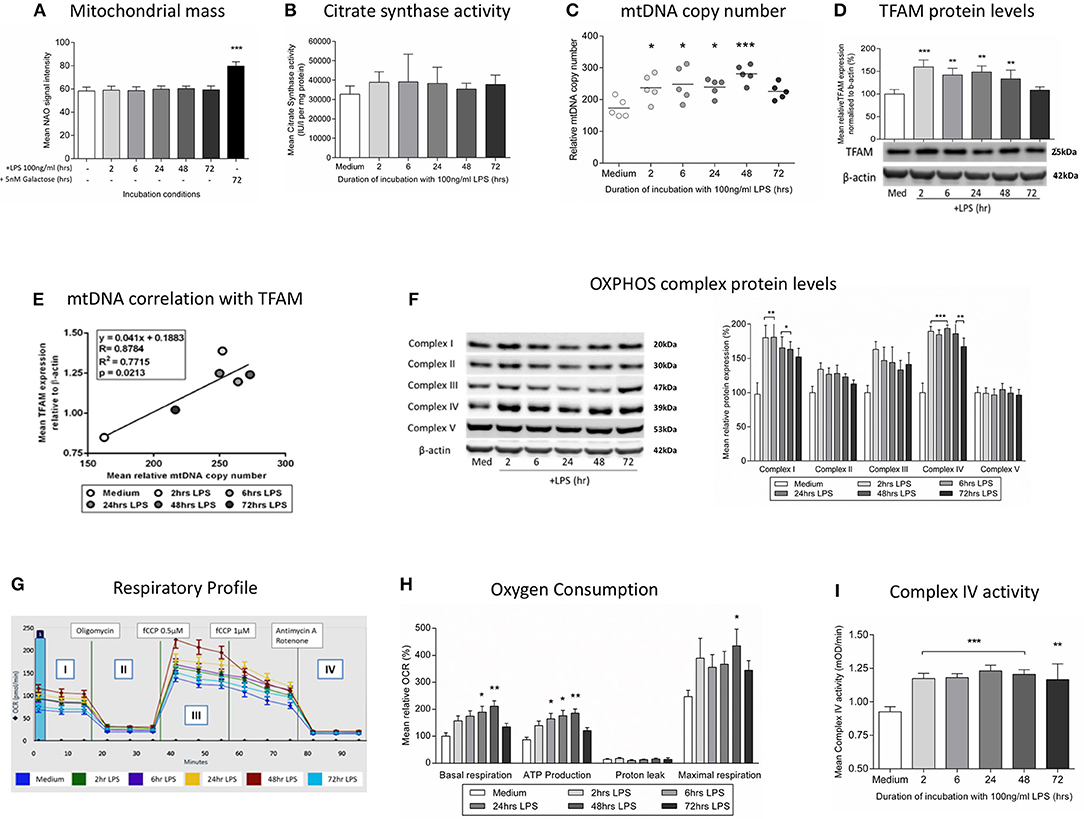
Frontiers | Exposure of Monocytic Cells to Lipopolysaccharide Induces  Coordinated Endotoxin Tolerance, Mitochondrial Biogenesis, Mitophagy, and  Antioxidant Defenses
Frontiers | Exposure of Monocytic Cells to Lipopolysaccharide Induces Coordinated Endotoxin Tolerance, Mitochondrial Biogenesis, Mitophagy, and Antioxidant Defenses

Macrophage Inflammatory Assay —BIO-PROTOCOL

Measurement of TLR4 and CD14 Receptor Endocytosis Using Flow Cytometry —BIO- PROTOCOL

Evidence for the Modulation of the Immune Response in Peripheral Blood  Mononuclear Cells after Stimulation with a High Molecular Weight β-glucan  Isolated from Lactobacillus fermentum Lf2 | bioRxiv
Evidence for the Modulation of the Immune Response in Peripheral Blood Mononuclear Cells after Stimulation with a High Molecular Weight β-glucan Isolated from Lactobacillus fermentum Lf2 | bioRxiv
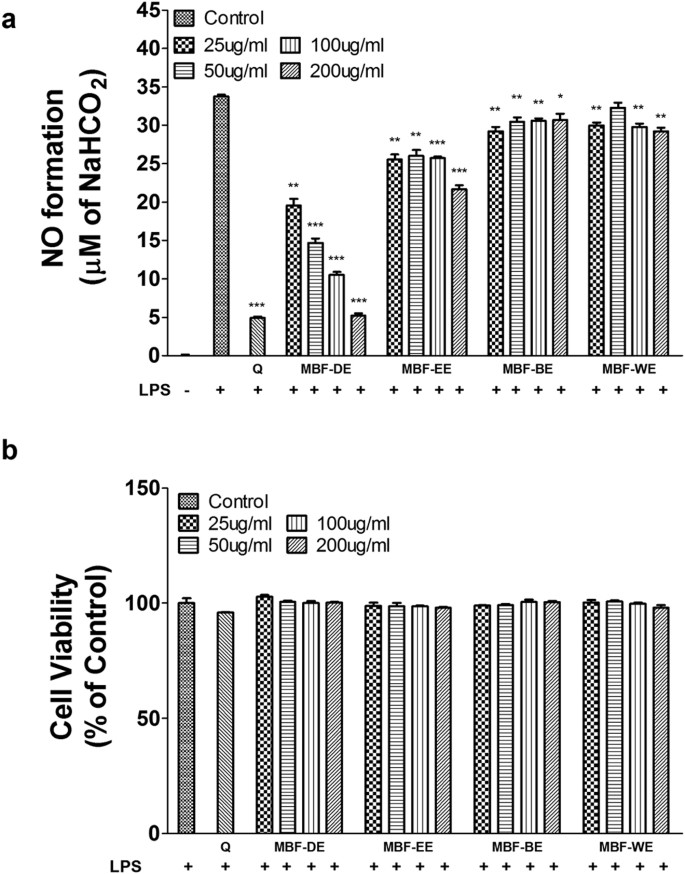
Mulberry fruit prevents LPS-induced NF-κB/pERK/MAPK signals in macrophages  and suppresses acute colitis and colorectal tumorigenesis in mice |  Scientific Reports
Mulberry fruit prevents LPS-induced NF-κB/pERK/MAPK signals in macrophages and suppresses acute colitis and colorectal tumorigenesis in mice | Scientific Reports

LPS stimulation of distinct subtypes of macrophages a Schematic... |  Download Scientific Diagram
LPS stimulation of distinct subtypes of macrophages a Schematic... | Download Scientific Diagram

Monocytes LPS stimulation protocol? | ResearchGate

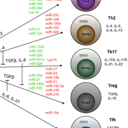
Lipopolysaccharide-driven Th2 Cytokine Production in Macrophages Is  Regulated by Both MyD88 and TRAM*
Lipopolysaccharide-driven Th2 Cytokine Production in Macrophages Is Regulated by Both MyD88 and TRAM*
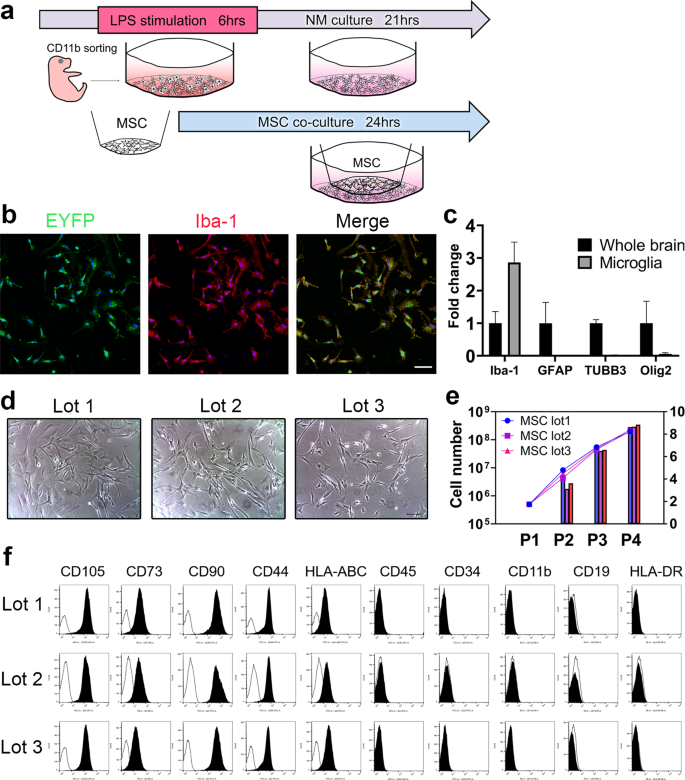
Umbilical cord-derived mesenchymal stromal cells immunomodulate and restore  actin dynamics and phagocytosis of LPS-activated microglia via PI3K/Akt/Rho  GTPase pathway | Cell Death Discovery
Umbilical cord-derived mesenchymal stromal cells immunomodulate and restore actin dynamics and phagocytosis of LPS-activated microglia via PI3K/Akt/Rho GTPase pathway | Cell Death Discovery

Standardized protocols for differentiation of THP-1 cells to macrophages  with distinct M(IFNγ+LPS), M(IL-4) and M(IL-10) phenotypes - ScienceDirect
Standardized protocols for differentiation of THP-1 cells to macrophages with distinct M(IFNγ+LPS), M(IL-4) and M(IL-10) phenotypes - ScienceDirect
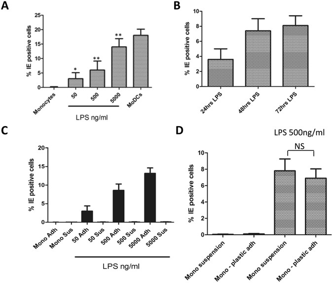
LPS promotes a monocyte phenotype permissive for human cytomegalovirus  immediate-early gene expression upon infection but not reactivation from  latency | Scientific Reports
LPS promotes a monocyte phenotype permissive for human cytomegalovirus immediate-early gene expression upon infection but not reactivation from latency | Scientific Reports
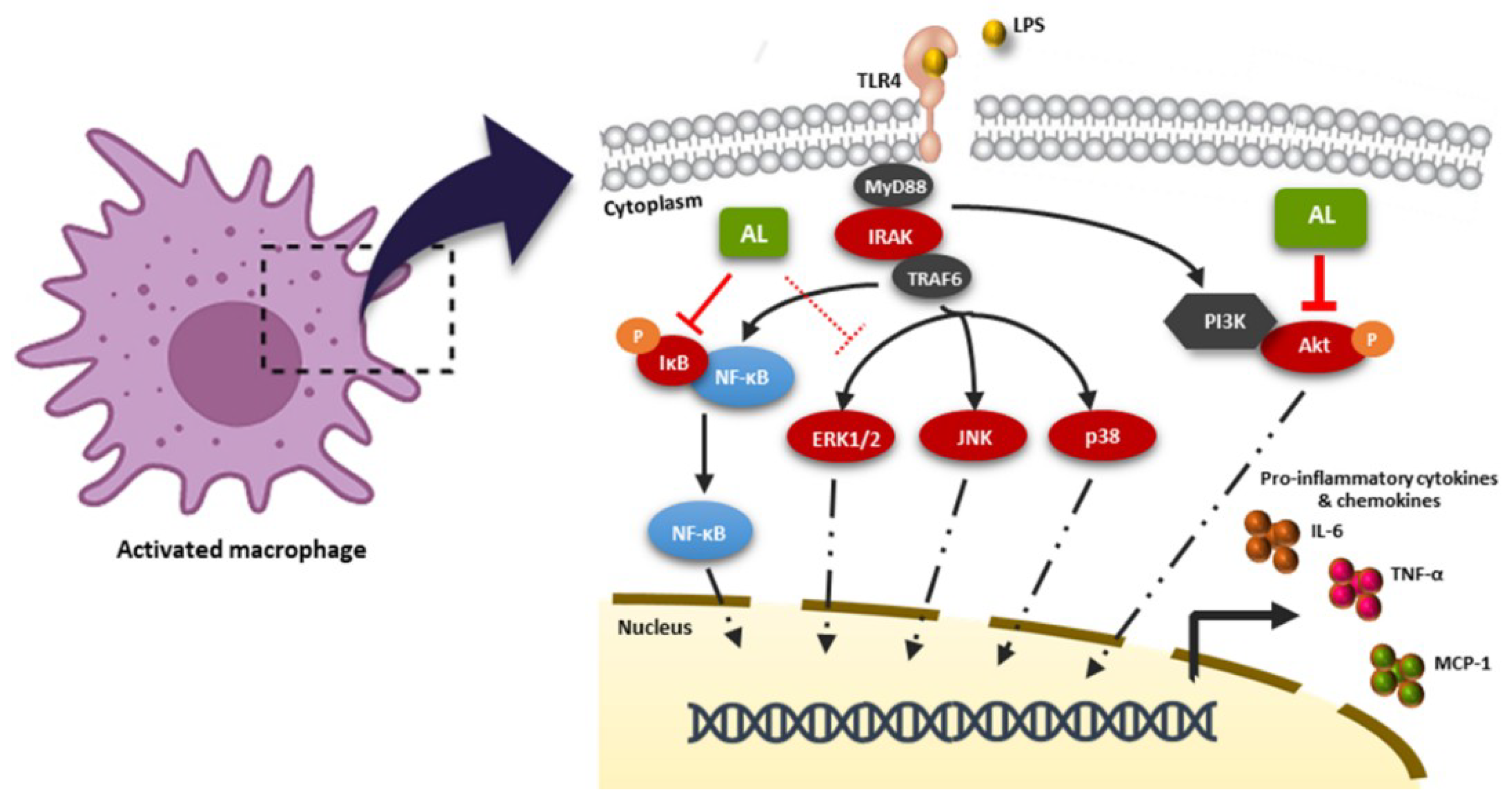
IJMS | Free Full-Text | Artocarpus lakoocha Extract Inhibits LPS-Induced  Inflammatory Response in RAW 264.7 Macrophage Cells
IJMS | Free Full-Text | Artocarpus lakoocha Extract Inhibits LPS-Induced Inflammatory Response in RAW 264.7 Macrophage Cells

Lipopolysaccharide-driven Th2 Cytokine Production in Macrophages Is  Regulated by Both MyD88 and TRAM*
Lipopolysaccharide-driven Th2 Cytokine Production in Macrophages Is Regulated by Both MyD88 and TRAM*
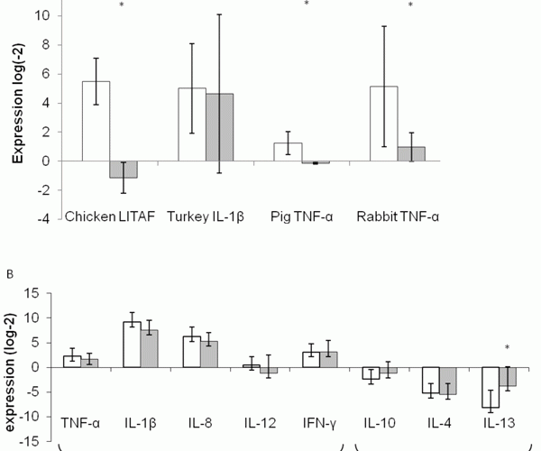
Ex-vivo LPS stimulation model coupled with quantitative PCR and its  multispecies application in immunonutrition - European Pharmaceutical Review
Ex-vivo LPS stimulation model coupled with quantitative PCR and its multispecies application in immunonutrition - European Pharmaceutical Review

LPS stimulation of PBMC suppresses SEB-induced cytokine production in T...  | Download Scientific Diagram
LPS stimulation of PBMC suppresses SEB-induced cytokine production in T... | Download Scientific Diagram

Frontiers | Exposure of Monocytic Cells to Lipopolysaccharide Induces  Coordinated Endotoxin Tolerance, Mitochondrial Biogenesis, Mitophagy, and  Antioxidant Defenses
Frontiers | Exposure of Monocytic Cells to Lipopolysaccharide Induces Coordinated Endotoxin Tolerance, Mitochondrial Biogenesis, Mitophagy, and Antioxidant Defenses

LPS‑induced proinflammatory cytokine expression in human airway epithelial  cells and macrophages via NF‑κB, STAT3 or AP‑1 activation
LPS‑induced proinflammatory cytokine expression in human airway epithelial cells and macrophages via NF‑κB, STAT3 or AP‑1 activation

LPS‑induced proinflammatory cytokine expression in human airway epithelial  cells and macrophages via NF‑κB, STAT3 or AP‑1 activation
LPS‑induced proinflammatory cytokine expression in human airway epithelial cells and macrophages via NF‑κB, STAT3 or AP‑1 activation